There are four main categories for neuromuscular diseases based on the affected site of the diseases:
(1)motor neurons
(2)peripheral nerve
(3)neuromuscular junction and muscle
(4)the diseases of muscle are in general coined myopathy.
Onset of neuromuscular disease can be acute, subacute and chronic. Depending on affected site, the clinical manifestations are widely different. The most common symptoms are muscle weakness, muscular atrophy and muscle pain.
A detailed clinical history and family history coped with careful physical examination are first step towards a correct diagnosis. Other investigations include blood tests, tests for suspected hereditary neuromuscular diseases, electromyography, MRI or muscle ultrasound and muscle & nerve biopsies.
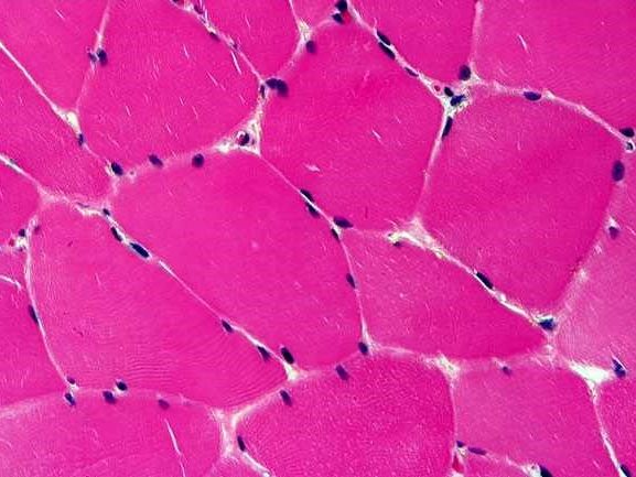
Muscle biopsy is a special procedure to precure a piece of muscle from diseased area. It assesses structural alternations in muscle fibers to provide evidence for diagnosis of neuromuscular diseases.
There are two biopsy methods:
(1)needle biopsy performed under localized anesthesia.
(2)open biopsy, which is conducted when a patient is under general anesthesia.
The choice of biopsy methods is usually based on clinical assessment. Needle biopsy can be used for various myositis, when there is inflammatory infiltrate in the muscle. Open biopsy is able to obtain a large piece of muscle about 1.0 cm in greatest dimension with good muscle fiber orientation for detailed microscopic and electron microscopic studies. While needle biopsy is often performed by neurologists at patient’s bedside, open biopsy is arranged by surgeons or orthopedic surgeons. Biopsy site is planned according to physical signs, electromyographic findings and MRI or muscle ultrasound features.
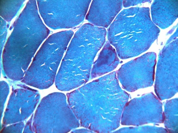
After the biopsy procedure, non-formalin fixed fresh muscle tissue is sending to histopathology laboratory for tissue processing, special muscle enzyme stains and even electron microscopy.
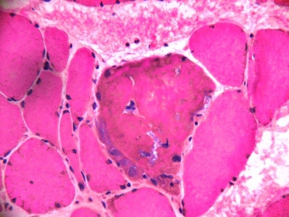
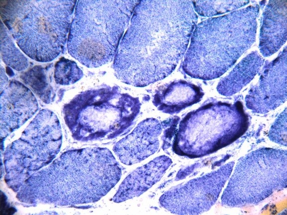
Before muscle biopsy is carried out, neurologists are going to order a series tests to rule out endocrine diseases, such as thyroid, adrenal dysfunction, and metabolic myopathy, such as Pompe disease. Muscle biopsy can provide evidence to confirm the diagnosis of myositis, i.e. polymyositis, dermatomyositis and toxic myopathy and vasculitis. It also helps the diagnosis of denervation and myopathies with structural abnormalities, like mitochondrial myopathy, lipid storage myopathy and some limb girdle dystrophy.
Nerve biopsy is to assist diagnosis of peripheral neuropathy. A proper consultation with neurologists is recommended before the biopsy. Microscopic examination of nerve sometimes may help the diagnosis of vasculitis, chronic inflammatory demyelinating polyneuropathy, amyloidosis, demyelination and axonal degeneration. Because of segmental involvement of the diseases, nerve biopsy is not always informative, hence it has limited diagnostic value. Nerve biopsy is an open biopsy. The biopsied nerve is mostly from sural nerve near the ankle joint.
Summary
Diagnosis of neuromuscular disease is a stepwise exercise following an algorithm, by which differential considerations are excluded or included. The diagnostic pathway involves a number of tests by multidisciplinary professionals, including clinical lab, imaging, electronic physiology and muscle/nerve biopsy. For some instances, the confirmatory diagnosis may take years to establish.